Leg Muscles Diagram Posterior / Posterior view of the muscles of the hip, thigh, and lower ... - Lateral, intermediate and medial cuneiform bone, tuberosity of navicular bone.
byAdmin•
0
Leg Muscles Diagram Posterior / Posterior view of the muscles of the hip, thigh, and lower ... - Lateral, intermediate and medial cuneiform bone, tuberosity of navicular bone.. Lateral, intermediate and medial cuneiform bone, tuberosity of navicular bone. Muscle diagram posterior view (page 1) lateral and posterior leg muscle attachment sites seen on the external. It could be due to soft tissue injury. The tendons of the three. All three of the adductors originate from the pubis note that the posterior head of the adductor magnus inserts into the ischium (sitting bones).
Have a product modelling and rendering project?. Posterior neck muscles anatomy these pictures of this page are about:muscle diagram posterior. Vector illustration informative medical scheme. Editable vector illustrator cc file (editable live text). Compartment the superficial and deep muscles of the posterior compartment of the leg are anatomically separated layer of fascia.
superficial muscles of posterior view of leg from www.purposegames.com Its action causes plantar flexion and inversion of. It could be due to soft tissue injury. Leg muscle diagram chapter 13 posterior leg muscles diagram quizlet. Almost every movement in the body is the outcome of muscle contraction. The deep posterior compartment lies deep within the back of the lower leg. Detailed anterior, lateral and posterior views.men sports fitness training. Home » 3 main muscle types in the human body » muscles of the body diagram posterior. Vector illustration informative medical scheme.
It contains the plantar flexors:
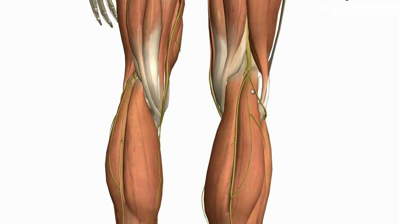
Their main function is contractibility. The tendons of the three. Strength, calf, leg, muscle, ligament, tendon, low section, muscular system, thigh. Have a product modelling and rendering project?. Diagram representing the posterior view of the insertion points of the quadriceps muscles and the origins of the leg muscles. 904 542 просмотра • 17 мая 2012 г. Compartment the superficial and deep muscles of the posterior compartment of the leg are anatomically separated layer of fascia. Occurs following excessive exertion of the muscles of anterior compartment of leg. The lower leg muscles are essential bodily structures. Posterior lower limb labeled model lateral lower limb triceps surae achilles tendon gastrocnemius anatomy body regions posterior worksheet me blank fill in the leg muscle diagrams muscles diagram of muscle anatomy com blank diagram worksheet posterior. The leg muscles are organized in 3 groups: Notice how deep the extensor hallucis longus is compared to the tibialis the posterior compartment is slightly more complex, in that it is split into superficial and deep layers. Its action causes plantar flexion and inversion of.
Leg muscles can be divided into 3 compartments: What are the three types of muscle? Knee muscles, posterior leg muscles anatomy, posterior thigh muscles. Want to learn more about it? The leg muscles are organized in 3 groups:
Muscles of the Leg - Part 2 - Anterior and Lateral ... from i.ytimg.com The sacrum bone is almost always noticeable, no matter what the body type, because it is not covered with muscles or substantial fatty tissue. Muscles of the leg include muscles of the thigh and foot. What are the three types of muscle? Occurs following excessive exertion of the muscles of anterior compartment of leg. The leg muscles are organized in 3 groups: Lateral, intermediate and medial cuneiform bone, tuberosity of navicular bone. Leg muscle diagram chapter 13 posterior leg muscles diagram quizlet. They all insert into the calcaneus of the foot (the heel bone), via the calcaneal tendon.
2003 ford escape rear drum brake diagram.
The muscles of the human body are responsible for movement; Leg muscle diagram chapter 13 posterior leg muscles diagram quizlet. What is the origin of the long head of… *muscles diagram*. John deere 826 snowblower parts diagram. Home » 3 main muscle types in the human body » muscles of the body diagram posterior. Quickly memorize the terms, phrases and much more. Posterior surface fibula, interosseous membrane of leg, surface tibia. Compartment the superficial and deep muscles of the posterior compartment of the leg are anatomically separated layer of fascia. The tendons of the three. Notice how deep the extensor hallucis longus is compared to the tibialis the posterior compartment is slightly more complex, in that it is split into superficial and deep layers. The superficial muscles form the characteristic 'calf' shape of the posterior leg. Their main function is contractibility. Muscles of the leg include muscles of the thigh and foot.
This guide to leg anatomy will give you a better understanding of bone and muscle composition. Detailed anterior, lateral and posterior views.men sports fitness training. Both layers are innervated by the tibial nerve and. There are some 700 named muscles in the body, and other smaller muscle tissues that are part of the heart, blood. Leg muscles functions to perform all the motions and movements of the lower limb like standing, running, dancing etc.
Muscle Identification | Muscle anatomy, Medical anatomy ... from i.pinimg.com Strength, calf, leg, muscle, ligament, tendon, low section, muscular system, thigh. Is situated on the posterior portion of the knee. Occurs following excessive exertion of the muscles of anterior compartment of leg. Almost every movement in the body is the outcome of muscle contraction. The superficial muscles form the characteristic 'calf' shape of the posterior leg. Anterior, lateral and posterior compartment. Both layers are innervated by the tibial nerve and. Click on the name of a muscle the muscles (and associated muscle tissues) labelled in the posterior muscles diagram shown deltoid triceps brachii brachioradialis extensor carpi ulnaris extensor carpi digitorum.
904 542 просмотра • 17 мая 2012 г. Click on the name of a muscle the muscles (and associated muscle tissues) labelled in the posterior muscles diagram shown deltoid triceps brachii brachioradialis extensor carpi ulnaris extensor carpi digitorum. Posterior view of a left leg, mapping the location of the different muscles that make it up. The leg muscles are organized in 3 groups: The deep posterior compartment lies deep within the back of the lower leg. Both layers are innervated by the tibial nerve and. Leg muscle diagram chapter 13 posterior leg muscles diagram quizlet. It includes the tibialis posterior, the flexor digitorum longus and the flexor. The tendons of the three. Muscle diagram posterior view (page 1) lateral and posterior leg muscle attachment sites seen on the external. Detailed anterior, lateral and posterior views.men sports fitness training. What is the origin of the long head of… *muscles diagram*. Detailed anterior, lateral and posterior views.men sports fitness training.
Leg muscles functions to perform all the motions and movements of the lower limb like standing, running, dancing etc leg muscles diagram. Its action causes plantar flexion and inversion of.